
An unusual cause of acute pulmonary embolism: giant hepatic hemangioma
Keywords:
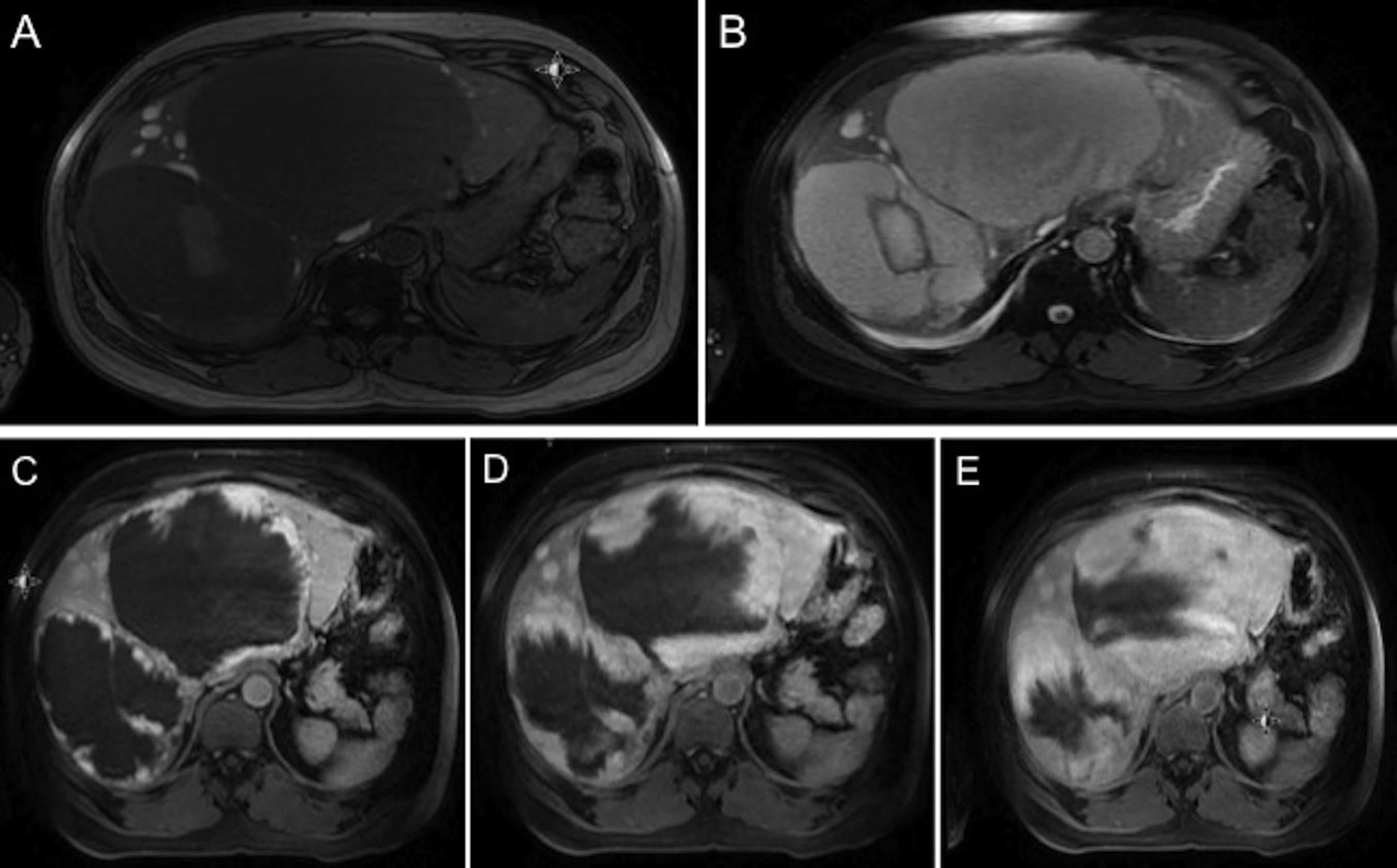
giant hepatic hemangioma, acute pulmonary embolism, inferior vena cava thrombosis
Abstract
Hemangiomas are the most common benign hepatic tumors and are usually asymptomatic. Lesions measuring more than 4 cm in diameter are known as “giant hemangiomas” and may cause various symptoms or complications depending on the size, the location, and the degree of compression of adjacent structures. Pulmonary embolism is a very rare complication of giant hepatic hemangiomas. In this case report, we describe a patient with acute pulmonary emboli, which presumably originated from laminar thrombi in the inferior vena cava caused by compression by giant hepatic hemangiomas.Downloads
Download data is not yet available.
Published
2016-06-17
How to Cite
Hatice Duygu Bas, H. D., Rassameehiran, S., Baser, K., Srisung, W., Bashir, M., & Woreta, T. (2016). An unusual cause of acute pulmonary embolism: giant hepatic hemangioma. The Southwest Respiratory and Critical Care Chronicles, 4(15), 66-69. Retrieved from https://pulmonarychronicles.com/index.php/pulmonarychronicles/article/view/277
Section
Case Reports


